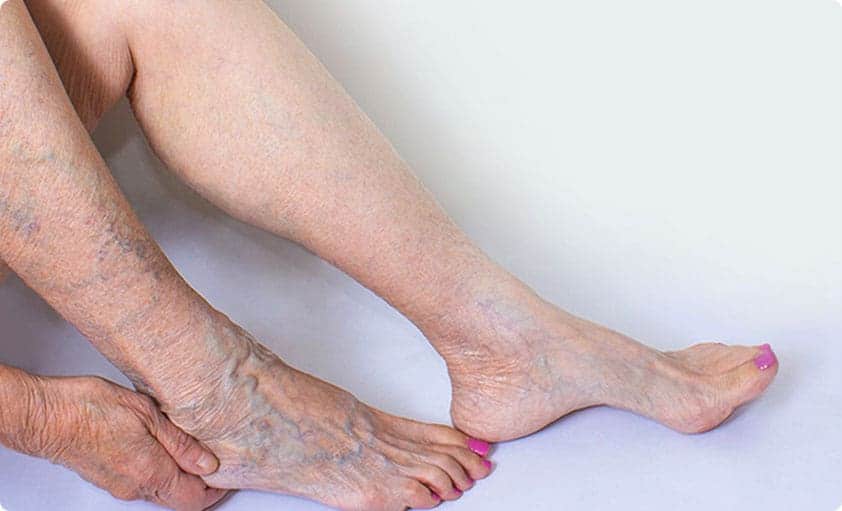
Varicose veins are veins that have become swollen, twisted, and visible through the skin. They typically develop in the legs, where the veins have to work against gravity to transport blood back to the heart. Normally, healthy veins possess valves that allow blood to only flow in one direction and prevent backflow. However, in varicose veins, these valves become weak or damaged, leading to accumulation of blood in the veins. This condition causes the veins to bulge and twist, exhibiting their characteristic appearance.
The symptoms of varicose veins can vary from mild to severe, common ones often include:
- Visible Veins: The most obvious sign is the appearance of dark blue or purple veins that are visibly bulging and twisted beneath the skin.
- Aching and Pain: Many individuals experience aching or discomfort, which can worsen after standing or sitting for extended periods.
- Swelling: Swelling, especially in the ankles and lower legs, can occur due to fluid buildup in the affected area.
- Itching and Burning: Some people may experience itching and a burning sensation around the veins.
- Leg Cramps: Varicose veins can lead to painful leg cramps, particularly at night.
Several factors can elevate the likelihood of developing varicose veins, including:
- Heredity: A family history of varicose veins increases the likelihood of developing them.
- Age: The risk of varicose veins increases with age, as vein walls naturally lose elasticity.
- Gender: Females have a higher probability of developing varicose veins because of hormonal shifts experienced during pregnancy and menopause.
- Obesity: Excess weight can put extra pressure on the veins, increasing the risk of varicose veins.
- Prolonged Standing or Sitting: Occupations or lifestyles that involve prolonged periods of sitting or standing may contribute to varicose veins.
To diagnose varicose veins, a healthcare professional will typically conduct a physical examination and ask about your symptoms and medical history. In certain instances, supplementary examinations, such as ultrasound, may be conducted to obtain a better view of the veins and evaluate blood flow.
The aim of varicose veins treatment is to alleviate symptoms and avert potential complications. Treatment options include:
Conservative Non-Surgical Treatment
1. Lifestyle Modifications: Simple lifestyle changes can help manage symptoms. These include regular exercise, elevating the legs, wearing compression stockings, and avoiding long periods of sitting or standing.
2. Compression Therapy: Tight compression stockings promote blood flow in the veins to alleviate some discomfort and prevent the progression of disease.
Minimally Invasive Procedures
1. Radiofrequency Ablation (RFA): RFA is a procedure which uses radiofrequency energy to heat and seal the vein.
2. Microwave Ablation (EMWA): EMWA is a procedure similar to RFA, but uses microwave energy to heat and seal the vein.
3. Venseal™/VenaBlock Glue Closure: A small quantity of specially formulated medical adhesive is applied to permanently seal the varicose vein. This sealing process redirects the blood to nearby healthy veins, offering relief.
4. Sclerotherapy: This minimally invasive procedure involves injecting a solution into the affected vein, which causes it to close and eventually be absorbed by the body.
Case Study 1
Patient presented with bilateral lower limb varicose veins, with left dorsal foot eczema and skin changes. After ultrasound examination, possible procedures including high-tie ligation, phlebectomies, radiofrequency ablation (RFA), and sclerotherapy, were discussed. The decision was then made to operate.
RFA and ClariVein ablation was performed on the left great saphenous vein. Under Ultrasound guidance, a radiofrequency electrode was inserted, and radiofrequency energy was used to heat the wall of the target vein, closing it off. Multiple phlebectomies via pinhole incisions were performed on the left leg, and segments of varicose veins were removed surgically. Bilateral sclerotherapy was also performed with sclerosing agents were injected into target veins in both legs to seal off residual veins.
Successful procedure and patient remains free from varicose veins 5 years post-procedure.
Case Study 2
Patient presented with bilateral lower limb varicose veins with thrombophlebitis (inflammation and swelling) of left medial calf and varicose veins. Left great saphenous vein had reflux from sapheno-femoral junction to the upper thigh which drains into the knee with more reflux from knee to ankle. 6 medial calf incompetent perforator veins were identified on ultrasound with drainage into medial calf varicosities. There were dilated reticular veins around the ankle and medial and lateral foot.
Endovenous ablation with microwave ablation under tumescence cover for the thigh segment and ClariVein for the knee to angle segment was performed on the left great saphenous vein via small pinhole punctures. Multiple phlebectomies to remove vein segments and ultrasound guided sclerotherapy were performed on both leg varicosities around ankles and the lower and lateral calf.
Patients symptoms resolved and she remains free from varicose veins 2 years post procedure.
Case Study 3
Patient presented with bilateral lower limb varicose veins, with varicosities present over thighs and calves. Thrombophlebitis was noted in the right medial gaiter region. The decision was made to operate.
Endovenous ablation with venaseal glue was used to treat the right great saphenous vein, closing it off. Multiple phlebectomies were then performed bilaterally, and the varicosities were avulsed via small needle stabs. Larger varicosities were treated by sclerotherapy under fluoroscopy guidance.
Swelling lessened, and extent of bruising was observed to have decreased.


