Deep vein thrombosis (DVT) is the spontaneous formation of blood clots in deep veins, primarily in the lower or upper limbs. Most cases are treatable with medication, but those with clots extending above the knee or upper arm face a higher risk of life-threatening complications, for example, a condition known as Pulmonary Embolism (PE). This condition is life-threatening as it prevents oxygen transfer to the venous blood and poses a significant risk of death.
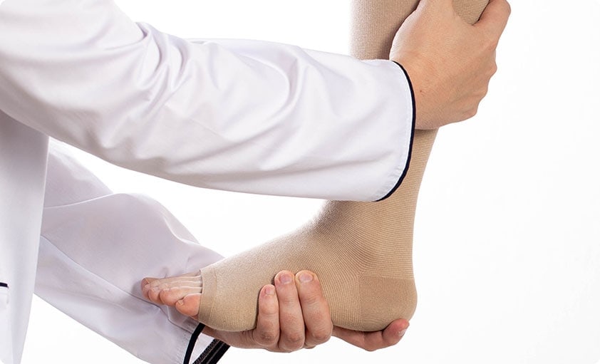
- Swollen and tender arms/legs
- Fast heartbeat
- Shortness of breath, especially when accompanied by limb swelling, may signal a pulmonary embolism and requires
- immediate medical attention
- Long periods of immobility
- Major surgeries
- Obesity
- Dehydration
- Genetic predisposition
- Underlying cancer
Diagnosis involves a risk factor assessment, ultrasound scans to detect clots, CT scans for pulmonary arteries, and ventilation-perfusion scans for suspected PE. Blood tests for pro-clotting factors and tumor markers may be conducted if cancer is suspected.
Pharmacological treatments for DVT:
Depending on health history, thinning blood medication (anti-coagulation) is prescribed to prevent blood clot formation, and dissolve existing blood clots for about six months.
Minimally invasive mechanical treatments for DVT:
If the DVT is extensive (e.g. extending up the thigh and into the pelvic veins), it is sometimes necessary to use a combination of mechanical devices and particular drugs to dissolve the clots. This minimally invasive process, known as thrombolysis, is often done if the DVT is less than two weeks old. The thrombolysis is a preventive measure for other long-term DVT complications including Post-Thrombotic Syndrome (PTS) and limb venous ulcers. Other additional treatments include the use of stents to keep the compressed veins open.
Case Study 1
A patient presented with right upper limb central vein thrombosis. As haemoglobin levels were low pre-procedure, the decision was made to use the Inari ClotTriever to reduce blood loss.
The Inari ClotTriever is a mechanical device specifically designed for optimal clot removal with minimal blood loss. In this procedure, a catheter was inserted into the vein, and the device was introduced, where it mechanically retrieved the clot, while preventing a further drop in haemoglobin levels. The clot was hence removed safely and efficiently, and damage to the surrounding tissue was minimised.
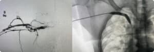
After surgery, right arm swelling decreased and a pulmonary embolus to the lung was averted.
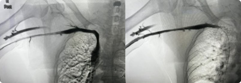
Case Study 2
Patient presented with an acute DVT extending from the tibial veins to the mid-superficial femoral vein. The patient experienced calf pain and aching despite taking oral anticoagulation medicine and compression stockings.
The left posterior tibial vein was accessed with an ultrasound guide and a wire was inserted into the femoral vein. Heparin was administered and thrombolysis was performed from the ankle to the superficial femoral vein with a combination of penumbra lighting 7 device and an angiojet device.
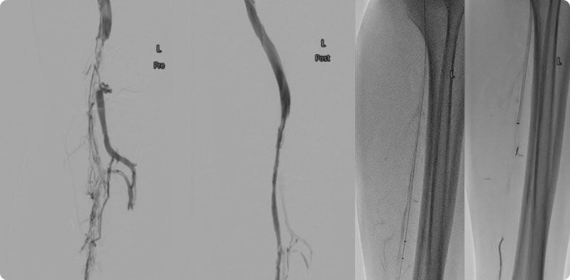
Angiogram showed return of patency with minimal clots left and good inflow via the peroneal and posterior tibial vein into the superficial femoral vein. Patient symptoms of leg pain and swelling resolved. He had to take oral anti-coagulation medication for months.


